Vegetos

(An International Journal of Plant Research & Biotechnology)
(eISSN: 2229-4473)
Exploring the potential of aqueous extract of Indian cherry fruit (Carissa carandas L.) as a natural plant histological stain
*Article not assigned to an issue yet
Short Communication | Published: 05 October, 2026
First Page: 0
Last Page: 0
Views: 30
Keywords: n Carissa stain, Dermal tissue system, Ground tissue system, Histological stain, Indian cherry
Abstract
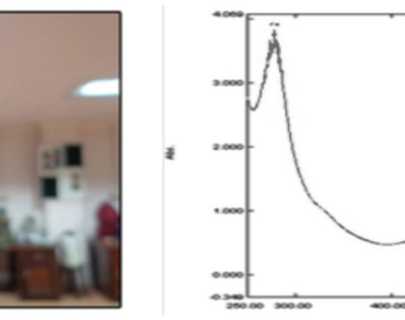
Natural dyes are eco-friendly, non-toxic, cost-effective and can be used for staining various plant tissues. We present our preparation of a natural stain from the aqueous extract of Carissa carandas fruits and its application for histological staining. We compared Carissa staining to safranin O staining of specimens of different plant tissues (Algae, Bryophytes, Pteridophytes, Gymnosperms and Angiosperms). The Carissa extract stain was red and exhibited absorption peaks at 277 and 511 nm. The pH of the Carissa extract stain was 3.2. The red color of the Carissa stain was due to its anthocyanin content. The Carissa extract stained pink colour on cuticle, epidermis, bulliform cells, pyrenoids, parenchyma, collenchyma, sclerenchyma, xylem and phloem tissues similar to safranin O. We found that the Carissa extract stains different types of plant tissues. We suggest that Carissa stain can be used as a natural histological stain for plant tissues.
References
Ahmed M, Zhang H, Xiong Y (2026) Anthocyanin structural types and stability: implications for natural colourant applications in food systems. Foods 15(12):2080. https://doi.org/10.3390/foods15122080
Crang R, Lyons-Sobaski S, Wise R (2018) Parenchyma, collenchyma, and sclerenchyma. Plant Anatomy. Springer, Cham. https://doi.org/10.1007/978-3-319-77315-5_6
Dang G, Li J, Yin C, Wang W, Zhang K et al (2024) Deciphering pectin: a comprehensive overview of its origins, processing, and promising utility. ACS Omega 10(1):1–15. https://doi.org/10.1021/acsomega.4c01843
Deepak MS, Omman P (2013) Use of dye extract of Melastoma malabathricum Linn. for plant anatomical staining. Act Biol Ind 2:456–460
Deepali K, Lalita S, Deepika M (2014) Application of aqueous plant extracts as biological stains. Int J Sci Eng Res 5(2):1586–1589
Dey P, Dey P, Hoque M, Rahman Md, Badhon B et al (2025) Sustainable and eco-friendly natural dyes: a holistic review on sources, extraction, and application prospects. Tex Res J 0(0):–1. https://doi.org/10.1177/00405175251321139
Dong W, Yang X, Zhang N, Chen P, Sun J, Harnly JM, Zhang M (2024) Study of UV–Vis molar absorptivity variation and quantitation of anthocyanins using molar relative response factor. Food Chem 444:138653. https://doi.org/10.1016/j.foodchem.2024.138653
Grigore MN, Toma C (2017) Bulliform cells. Anatomical adaptations in halophytes. Springer Int pub Cham Switz 325–338. https://doi.org/10.1007/978-3-319-66480-4_8
Hoffman LR (1968) Observations on the fine structure of oedogonium IV. The mature pyrenoid of Oe. cardiacum. Trans Am Microsc Soc 87(2):178–185. https://doi.org/10.2307/3224438
Jan HU, Shinwari ZK, Khan AA (2011) Staining effect of dye extracted from dry leaves of Lawsonia inermis Linn. (henna) on angiospermic stem tissue. Pak J Bot 43(1):383–389
Jasphin S, Raghavendra AP, Solomon MC, Amuthan A et al (2023a) Anthocyanins: a hue for histology—systematic review. Malay J Sci 42(1):106–125. https://doi.org/10.22452/mjs.vol42no1.10
Jarvis MC (2012) Sclerenchyma. In: eLS, (Ed.). https://doi.org/10.1002/9780470015902.a0002082.pub2
Jasphin S, Parampalli AR, Solomon MC, Amuthan A, Singh MK, Kairanna NK (2023b) Anthocyanins: a hue for histology—Systematic Review. Mal J Sci 42(1):106–125. https://doi.org/10.22452/mjs.vol42no1.10
Lago E, Vasconcelos A, Araujo A, Espindola L, Quelemes P et al (2025) Natural pigments as eco-friendly staining agents: evaluation of β-carotene-rich carrot and betanin-rich beetroot extracts for bacterial biofilm visualisation. Nat Prod Res 1–5. https://doi.org/10.1080/14786419.2025.2584437
Long BM, Matsuda Y, Moroney JV (2024) Algal chloroplast pyrenoids: evidence for convergent evolution. Proc Natl Acad Sci 121(14):e2402546121. https://doi.org/10.1073/pnas.2402546121
Lux A, Morita S, Abe J, Ito K (2005) An improved method for clearing and staining free-hand sections and whole-mount samples. Ann Bot 96(6):989–996. https://doi.org/10.1093/aob/mci266
Mokshina N, Sautkina O, Gorshkov O, Mikshina P (2025) A fresh look at celery collenchyma and parenchyma cell walls through a combination of biochemical, histochemical, and transcriptomic analyses. Int J Mol Sci 26(2):738. https://doi.org/10.3390/ijms26020738
Raj KB, Chandrashekaraiah P, Kalaivani P, Oza N, Ramadoss K et al (2026) Syzygium cumini (jamun) as a natural histological stain: a sustainable alternative to hematoxylin and eosin. J Oral Res Rev 18(1):42–50. https://doi.org/10.4103/jorr.jorr_78_25
Sachdev SS, Chettiankandy TJ, Sonawane SG, Sardar MA, Kende PP, Pakhmode V (2021) Toward developing natural histologic stains using anthocyanins: a novel approach. J Oral Maxillofacial Pathol 25(1):199–200
Sarkar R, Kundu A, Banerjee K, Saha S (2018) Anthocyanin composition and potential bioactivity of karonda (Carissa carandas L.) fruit: an indian source of biocolorant. LWT food Sci Tech 93:673–678. https://doi.org/10.1016/j.lwt.2018.04.012
Shahidi F, Athiyappan KD (2025) Polyphenol-polysaccharide interactions: molecular mechanisms and potential applications in food systems: a comprehensive review. Food Prod Process Nutr 7(1):42. https://doi.org/10.1186/s43014-025-00322-3
Shahidi F, Dissanayaka CS (2023) Phenolic-protein interactions: insight from in-silico analyses: a review. Food Prod Process Nutr 5(2):1–21. https://doi.org/10.1186/s43014-022-00121-0
Shi C, Guo C, Wang S, Li W, Zhang X, Lu S, Ning C, Tan C (2024) The mechanism of pectin in improving anthocyanin stability and the application progress of their complexes: A review. Food Chem 24:101955. https://doi.org/10.1016/j.fochx.2024.101955
Wagiyanti H, Noor R (2017) Red dragon fruit (Hylocereus costaricensis Britt. Et R) as a natural staining alternative in microscopic observation of the plant tissues: the practical guide in senior high school. J Pendidikan Biol Ind 3:232–237. https://doi.org/10.22219/jpbi
Author Information
Department of Botany, St. Teresa’s College (Autonomous), Cochin, India