Vegetos

(An International Journal of Plant Research & Biotechnology)
(eISSN: 2229-4473)
In vitro propagation, in vitro flowering, ex vitro root regeneration and foliar micro-morphological analysis of Hedyotis biflora (Linn.) Lam
Revathi J., Manokari M., Latha R., Priyadharshini S., Kher Mafatlal M., Shekhawat Mahipal S.
Research Articles | Published: 23 October, 2019
First Page: 609
Last Page: 619
Views: 4288
Keywords: In vitro flowering, Ex vitro rooting, Hedyotis biflora , Micro-morphological studies
Abstract
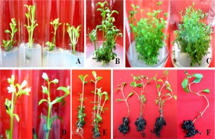
Hedyotis biflora (Linn.) Lam is an important herb, used in the Indian and Chinese traditional systems of medicines. Indiscriminate harvesting of this plant resulted in decline of its population from the natural habitats. The present study reports effective micropropagation and in vitro flowering in H. biflora. Healthy plants were maintained in the greenhouse to collect fresh explants (nodal segments). Average 3.2 shoot bud/node with 88% shoot regeneration obtained on Murashige and Skoog’s (MS) medium with 2.0 mg L−1 6-benzylaminopurine (BAP) and additives like 50 mg L−1 of ascorbic acid and 25 mg L−1 each of arginine, adenine sulphate and citric acid. About 84.5 shoots, with 8.1 cm shoot length were obtained on MS basal medium supplemented with 1.0 mg L−1 BAP, 0.5 mg L−1 kinetin (Kin) and 0.1 mg L−1 indole-3 acetic acid (IAA) and additives. In vitro flowers were induced in 91% cultures (6.8 flowers per shoot) when the shoots were cultured on MS medium supplemented with 1.5 mg L−1 BAP and 0.1 mg L−1 IAA at 12-h photoperiods. In vitro raised shoots were rooted with the help of indole-3 butyric acid (IBA) via in vitro and ex vitro techniques. The micropropagated plants were acclimatized with 91% survival rate in the field. Upon transfer of plantlets from the in vitro to ex vitro environments, there were significant changes in stomata, vein-islets and raphids. These micro-morphological changes may help micropropagated plantlets to establish under the field conditions. The present protocol is suitable for large scale micropropagation, conservation and commercial production of this plant. The protocol of in vitro flowering could be useful in understanding the in vitro pollination biology of H. biflora.
References
- Behera SK, Rajasekaran C, Payas S et al (2018) In vitro flowering in Oldenlandia umbellata L. J Ayurveda Integr Med 9:99–103. https://doi.org/10.1016/j.jaim.2017.02.011
- Brar J, Shafi A, Sood P et al (2014) In vitro propagation, biochemical studies and assessment of clonal fidelity through molecular markers in Bambusa balcooa. J Trop For Sci 26:115–124
- Casson S, Gray JE (2008) Influence of environmental factors on stomatal development. New Phytol 178:9–23. https://doi.org/10.1111/j.1469-8137.2007.02351.x
- Chiu T-C, Chang C (2018) In vitro flowering and breeding of Erycina pusilla. In: Lee Y-L, Yeung EC-T (eds) Orchid propagation: from laboratories to greenhouses—methods and protocols. Springer Science + Business Media, New York, pp 257–265
- Corbineau F, Come D (1980) Some particularities of the germination of Oldenlandia corymbosa L. seeds. Isr J Bot 29:157–167
- Croxdale JL (2000) Stomatal patterning in angiosperms. Am J Bot 87:1069–1080. https://doi.org/10.2307/2656643
- Datta PC, Sen A (1969) Pharmacognosy of Oldenlandia corymbosa Linn. Q J Crude Drug Res 9:1365–1371. https://doi.org/10.3109/13880206909066279
- Ding X, Bai D, Qian J (2014) Novel cyclotides from Hedyotis biflora inhibit proliferation and migration of pancreatic cancer cell in vitro and in vivo. Med Chem Res 23:1406–1413. https://doi.org/10.1007/s00044-013-0746-6
- Ďurkovič J, Lengyelová A, Čaňová I et al (2009) Photosynthetic performance and stomatal characteristics during ex vitro acclimatisation of true service tree (Sorbus domestica L.). J Hortic Sci Biotechnol 84:223–227. https://doi.org/10.1080/14620316.2009.11512508
- e-Flora of China (2019) Hedyotis biflora (Linnaeus) Lamarck, Tabl. Encycl. 1: 272. 1792. http://www.efloras.org/florataxon.aspx?flora_id=2&taxon_id=200022098. Accessed 29 Mar 2019
- Franceschi VR, Nakata PA (2005) Calcium oxalate in plants: formation and function. Annu Rev Plant Biol 56:41–71. https://doi.org/10.1146/annurev.arplant.56.032604.144106
- Fukuda H, Ohashi-Ito K (2019) Vascular tissue development in plants. In: Grossniklaus U (ed) Current topics in developmental biology, vol 131. Academic Press + Elsevier Inc, Cambridge, pp 141–160
- Gamble JS (1915) Flora of the Presidency of Madras, vol 2. Newman and Adlard Publishers, London
- Gentry HS, Sauck JR (1978) The stomatal complex in Agave: groups Deserticolae, Campaniflorae, Umbelliflorae. Proc Calif Acad Sci 41:371–387
- González-Rodríguez A, Arias DM, Valencia S, Oyama K (2004) Morphological and RAPD analysis of hybridization between Quercus affinis and Q. laurina (fagaceae), two Mexican red oaks. Am J Bot 91:401–409. https://doi.org/10.3732/ajb.91.3.401
- Guo X, Wang RJ, Simmons MP et al (2013) Phylogeny of the Asian Hedyotis-Oldenlandia complex (Spermacoceae, Rubiaceae): evidence for high levels of polyphyly and the parallel evolution of diplophragmous capsules. Mol Phylogenet Evol 67:110–122. https://doi.org/10.1016/j.ympev.2013.01.006
- Hamzah AS, Jasmani H, Ahmad R, Baba AR (1997) New Anthraquinones from the roots of Hedyotis dichotoma. J Nat Prod 60:36–37. https://doi.org/10.1021/np960583
- Hanley ME, Lamont BB, Fairbanks MM, Rafferty CM (2007) Plant structural traits and their role in anti-herbivore defence. Perspect Plant Ecol Evol Syst 8:157–178. https://doi.org/10.1016/j.ppees.2007.01.001
- Hazarika BN (2003) Acclimatization of tissue-cultured plants. Curr Sci 85:1704–1712
- Hazarika BN (2006) Morpho-physiological disorders in in vitro culture of plants. Sci Hortic 108:105–120. https://doi.org/10.1016/j.scienta.2006.01.038
- Hernandéz-Valencia REM, López-Franco R, Ruíz-Ordoñez J et al (2003) The stomatal complex and epicuticular characteristics of crown rot disease of Agave tequilana Weber (Agavaceae). Acta Hortic 618:427–433. https://doi.org/10.17660/ActaHortic.2003.618.51
- Hickey LJ, Wolfe JA (1975) The bases of angiosperm phylogeny: vegetative morphology. Ann Missouri Bot Gard 62:538–589. https://doi.org/10.2307/2395267
- Ivanova M, Van Staden J (2010) Natural ventilation effectively reduces hyperhydricity in shoot cultures of Aloe polyphylla Schönland ex Pillans. Plant Growth Regul 60:143–150. https://doi.org/10.1007/s10725-009-9430-8
- João PRM, Veerle V, Moacir P, Maurice DP (2016) Physiological responses by Billbergia zebrina (Bromeliaceae) when grown under controlled microenvironmental conditions. African J Biotechnol 15:1952–1961. https://doi.org/10.5897/AJB2016.15584
- Johansen DA (1940) Plant microtechnique, edn 1. McGraw Hill Book Co, New York
- John CK, Nadgauda RS (1999) Review in vitro-induced flowering in bamboos. Vitro Cell Dev Biol Plant 35:309–315. https://doi.org/10.1007/s11627-999-0040-y
- Karthi C, Velayutham P (2016) In vitro organogenesis and rapid multiplication of Oldenlandia biflora L.—a little known medicinal plant. Int J Recent Sci Res 7:12434–12439
- Kostman TA, Tarlyn NM, Loewus FA, Franceschi VR (2001) Biosynthesis of L-ascorbic acid and conversion of carbons 1 and 2 of L-ascorbic acid to oxalic acid occurs within individual calcium oxalate crystal idioblasts. Plant Physiol 125:634–640. https://doi.org/10.1104/pp.125.2.634
- Kraus JE, Arduin M (1997) Manual básico de métodos em morfologia vegetal. Universidade Federal Rural do Rio de Janeiro, Rio de JaneiroEd. Universidade Federal Rural do Rio de Janeiro, Rio de Janeiro, Seropédica, RJ, Brasil
- Kumar K, Rao IU (2012) Morphophysiologicals problems in acclimatization of micropropagated plants in–ex vitro conditions—a reviews. J Ornam Hortic Plants 2:271–283
- Kumari A, Baskaran P, Van Staden J (2017) In vitro regeneration of Begonia homonyma—a threatened plant. South African J Bot 109:174–177. https://doi.org/10.1016/j.sajb.2016.12.027
- Leshem B (1983) Growth of carnation meristems in vitro: anatomical structure of abnormal plantlets and the effect of agar concentration in the medium on their formation. Ann Bot 52:413–415. https://doi.org/10.1093/oxfordjournals.aob.a086591
- Lodha D, Patel AK, Shekhawat NS (2015) A high-frequency in vitro multiplication, micromorphological studies and ex vitro rooting of Cadaba fruticosa (L.) Druce (Bahuguni): a multipurpose endangered medicinal shrub. Physiol Mol Biol Plants 21:407–415. https://doi.org/10.1007/s12298-015-0310-6
- Luna CV, Gonzalez AM, Mroginski LA, Sansberro PA (2017) Anatomical and histological features of Ilex paraguariensis leaves under different in vitro shoot culture systems. Plant Cell Tissue Organ Cult 129:457–467. https://doi.org/10.1007/s11240-017-1191-x
- Monje PV, Baran EJ (2002) Characterization of calcium oxalates generated as biominerals in Cacti. Plant Physiol 128:707–713. https://doi.org/10.1104/pp.010630
- Murashige T, Skoog F (1962) A revised medium for rapid growth and bio assays with tobacco tissue cultures. Physiol Plant 15:473–497. https://doi.org/10.1111/j.1399-3054.1962.tb08052.x
- Nakata PA (2012) Plant calcium oxalate crystal formation, function, and its impact on human health. Front Biol (Beijing) 7:254–266. https://doi.org/10.1007/s11515-012-1224-0
- Nimal Christhudas IVS, Praveen Kumar P, Sunil C et al (2013) In vitro studies on α-glucosidase inhibition, antioxidant and free radical scavenging activities of Hedyotis biflora L. Food Chem 138:1689–1695. https://doi.org/10.1016/j.foodchem.2012.11.051
- Panigrahi J, Dholu P, Shah TJ, Gantait S (2018) Silver nitrate-induced in vitro shoot multiplication and precocious flowering in Catharanthus roseus (L.) G. Don, a rich source of terpenoid indole alkaloids. Plant Cell Tissue Organ Cult 132:579–584. https://doi.org/10.1007/s11240-017-1351-z
- Pospíšilová J, Tichá I, Kadlechek P et al (1999) Acclimatization of micropropagated plants to ex vitro conditions. Biol Plant 42:481–497. https://doi.org/10.1023/A:1002688208758
- Prabhakar M (2004) Structure, delimitation, nomenclature and classification of stomata. Acta Bot Sin 46:242–252
- Qi Z, Pi E, Zhang X et al (2017) Ontogenesis of D-type stomata and cork-warts on the leaf epidermis of Camellia japonica (Theaceae) and functional assessment. Flora 228:24–30. https://doi.org/10.1016/j.flora.2017.01.010
- Revathi J, Manokari M, Shekhawat MS (2018) Optimization of factors affecting in vitro regeneration, flowering, ex vitro rooting and foliar micromorphological studies of Oldenlandia corymbosa L.: a multipotent herb. Plant Cell Tissue Organ Cult 134:1–13. https://doi.org/10.1007/s11240-018-1395-8
- Salisbury EJ (1932) The interrelations of soil, climate and organisms and the use of stomatal frequency as an integrating index of the water relation of the plant. Beih Bot Zbl 99:402–420
- Saranya Krishnan SR, Siril EA (2017) Enhanced in vitro shoot regeneration in Oldenlandia umbellata L. by using quercetin: a naturally occurring auxin-transport inhibitor. Proc Natl Acad Sci India Sect B Biol Sci 87:899–904. https://doi.org/10.1007/s40011-015-0672-0
- Sarasan V (2010) Importance of in vitro technology to future conservation programmes worldwide. Kew Bull 65:549–554. https://doi.org/10.1007/s12225-011-9250-7
- Shekhawat MS, Manokari M (2016) In vitro propagation, micromorphological studies and ex vitro rooting of cannon ball tree (Couroupita guianensis aubl.): a multipurpose threatened species. Physiol Mol Biol Plants 22:131–142. https://doi.org/10.1007/s12298-015-0335-x
- Shekhawat MS, Manokari M (2017) Comparative foliar micromorphological studies of in vitro and field transferred plants of Morinda citrifolia. Acta Bot Hung 59:427–438. https://doi.org/10.1556/034.59.2017.3-4.8
- Shekhawat MS, Kannan N, Manokari M (2015) In vitro propagation of traditional medicinal and dye yielding plant Morinda coreia Buch.–Ham. South African J Bot 100:43–50. https://doi.org/10.1016/j.sajb.2015.05.018
- Shekhawat MS, Manokari M, Kannan N (2017) Micromorphological response towards altered environmental conditions in subsequent stages of in vitro propagation of Morinda coreia. Environ Exp Biol 15:37–46. https://doi.org/10.22364/eeb.15.06
- Shin K-S, Park S-Y, Paek K-Y (2014) Physiological and biochemical changes during acclimatization in a Doritaenopsis hybrid cultivated in different microenvironments in vitro. Environ Exp Bot 100:26–33. https://doi.org/10.1016/j.envexpbot.2013.12.004
- Sivapraksam SSK, Karunakaran K, Subburaya U et al (2014) A review on phytochemical and pharmacological profile of Hedyotis corymbosa Linn. Int J Pharm Sci Rev Res 26:320–324
- Sridhar N, Kumar NVSP, Sasidhar D et al (2012) Antibacterial and phytochemical evaluation of Oldenlandia biflora L. and Pergularia daemia. Int J Drug Dev Res 4:148–152
- Su YH, Zhang XS (2014) The hormonal control of regeneration in plants. Curr Top Dev Biol 108:35–69
- Teixeira da Silva JA, Kerbauy GB, Zeng S et al (2014a) In vitro flowering of orchids. Crit Rev Biotechnol 34:56–76. https://doi.org/10.3109/07388551.2013.807219
- Teixeira da Silva JA, Zeng S, Cardoso JC et al (2014b) In vitro flowering of Dendrobium. Plant Cell Tissue Organ Cult 119:447–456. https://doi.org/10.1007/s11240-014-0561-x
- Teixeira da Silva JA, Hossain MM, Sharma M et al (2017) Acclimatization of in vitro-derived Dendrobium. Hortic Plant J 3:110–124. https://doi.org/10.1016/j.hpj.2017.07.009
- Ticha I, Radochova B, Kadlecek P (1999) Stomatal morphology during acclimatization of tobacco plantlets to ex vitro conditions. Biol Plant 42:469–474. https://doi.org/10.1023/A:1002450210939
- Wang D, Li X, Cheng Z, Long C (2018) In vitro preservation and micropropagation of Oreocharis mileense (W.T. Wang) M. Möller & A. Weber (Gesneriaceae) through shoot organogenesis. Vitro Cell Dev Biol Plant 54:606–611. https://doi.org/10.1007/s11627-018-9941-y
- Wong CTT, Taichi M, Nishio H et al (2011) Optimal oxidative folding of the novel antimicrobial cyclotide from Hedyotis biflora requires high alcohol concentrations. Biochemistry 50:7275–7283. https://doi.org/10.1021/bi2007004
- Wu C-C, Huang H-C, Kuo Y-H et al (2006) New cytotoxic 6-oxygenated 8,9-dihydrofurocoumarins, hedyotiscone A—C, from Hedyotis biflora. Planta Med 72:75–78. https://doi.org/10.1055/s-2005-873178
- Yuan Q, Zhao J, Yang J, Li L (2001) Survey on triterpenoids from Hedyotis and their spectroscopic characteristics. Zhongcaoyao 32:754–756
Author Information
Department of Botany, Kanchi Mamunivar Center for Postgraduate Studies, Puducherry, India