Histochemical, phytochemical, and cytotoxicity of Gyrinops versteegii “Madu” (Thymelaeaceae) against bacteria infectious
*Article not assigned to an issue yet
Mulyaningsih Tri, Febrianti Via, Sunarwidhi L. A., Muspiah Aida, Hidayati Ernin, Sukenti Kurniasih, Kurnianingsih Rina, Listiana Baiq Erna, Ito M., Yamada I.
Research Articles | Published: 10 July, 2025
First Page: 0
Last Page: 0
Views: 71
Keywords: n Gyrinops versteegiin , Agarwood, Histochemistry, Antibacterial, Lombok
Abstract
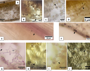
Gyrinops versteegii “Madu” is from Lombok Island, Indonesia. This plant contains metabolic compounds that have medicinal properties. This study aims to determine the histochemistry, and phytochemistry of the vegetative and reproductive organs of G. versteegii “Madu”, and also its cytotoxicity against 3 species of infectious bacteria (Escherichia coli, Pseudomonas aeruginosa and Proteus mirabilis). This research used a descriptive method which includes three activities. Histochemical preparations were made by hand free section, vegetative organs (wood, healthy bark, lichen bark, leaves) and reproductive organs (petals, stalks, fruit peels, and seeds), to detect metabolic compounds; The acetone extract for each organ was prepared and identify metabolic compounds was detected. The acetone extract tested on pure cultures of 3 species of infectious bacteria, with acetone as a negative control, Gentamycin Sulfate as a positive control, and each treatment was repeated 3 times. The results showed that the vegetative and reproductive organs contain 11 kinds of metabolites. Except the saponins and sesquiterpene lactones were not detected in the lamina. Ca-oxalate crystals and sesquiterpene lactones were not found in the seed. All organs acetone extract contained all the tested metabolic compounds, except for saponin compounds, but the stem bark associated with lichen was detected these compounds. The acetone extract of the flowers, have a higher inhibition than other extracts against E. coli, P. mirabilis and P. aeruginosa.

References
Andary, C., Longpierre, D., Cong, K., Hul, S., Zaremski, A. & Michaloud, G. A. (2019). Study of a chemotaxonomic marker to identify the genus Aquilaria (Thymelaeaceae). Bois et Forêts Des Tropiques, 341(3), 29–38.
Armbruster, C. E., Mobley, H. T., & Pearson, M. M. (2018). Pathogenesis of Proteus mirabilis Infection. EcoSal Plus, 8(1), 10.1128/ecosalplus.ESP-0009-2017.
Badria F.A., & Aboelmataaty W.S. (2019). Plant histochemistry: a versatile and indispensible tool in localization of gene expression, enzymes, cytokines, secondary metabolites and detection of plants infection and pollution. ACTA Scientific Pharmaceutical Sciences, 3(7), 88–100.
Budayanti, N., Aisyah, D., Fatmawati, N. N.., Tarini, N. M., Kozlakidis, Z., & Adisasmito, W. (2020). Identification and distribution of pathogens in a mayor tertiary hospital of Indonesia. Front. Public Health, 7(395), 1–8.
Dash, M., Patra, J. K., & Panda, P. P. (2008). Phytochemical and antimicrobial screening of extracts of Aquilaria agallocha Roxb. African Journal of Biotechnology., 7(20), 3531–3534. https://doi.org/10.5897/AJB08.623
Evans, W. C. (2009). Trease and Evans pharmacognosy. (16th ed.). Saunders Elsevier
Gardner, R. O. (1975). Vanillin-hydrochloric acid as a histochemical test for tannin. Stain-Technology, 50(5), 315–317.
Gaykhe, R. C., & Kadam, V. B. (2008). Hi. African Journal of Biotechnology. https://doi.org/10.5897/AJB08.623
Hashim, Y. Z. H.-Y., N, Kerr, P. G., Abbas, P., & Salleh, H. M. (2016). Aquilaria spp. (agarwood) as source of health beneficial compounds: A review of traditional use, phytochemistry and pharmacology. Journal of Ethnopharmacology, 189, 331–360. https://doi.org/10.1016/j.jep.2016.06.055
Hidayati, E., Handayani, & Sudharma, I.Md. (2023). Antibacterial activity of gyrinops versteegii fruit extracts against staphylococcus aureus and escherichia coli and GC-MS Analysis. Journal of Mathematical and Fundamental Sciences., 54(2), 249–260.
Jachula, J., Konarska, A., & Denisow, B. (2018). Micromorphological and histochemical attributes of flowers and floral reward in linaria vulgaris (plantaginaceae). Protoplasma, 255, 1763–1776.
Jaeger, R., & Cuny, E. (2016). Terpenoids with special pharmacological significance: a review. Natural Product Communications., 11(9), 1373–1390.
Jensen, W. A. (1962). Botanical Histochemistry: Principles and Practices. W.H. Freeman and company.
Johansen, D. A. (1940). Plant Microtechnique (first edit). McGraw-Hill Book Company, Inc.
Kadam, V. S., Fatima, S., Tambe, S. S., & Momin, R. K. (2013). Histochemical investigation of different organs of two medicinal. International Journal of Pharmaceutical Research and Bioscience, 2(4), 194–201.
Kadam V.B. (1999). Histochemical investigations of different organs of three endangered medicinal taxa of South Gujarat Forests. Journal Phytological Research, 12(1–2), 109–112.
Kotzé, M., & Eloff, J. N. (2002). Extraction of antibacterial compounds from combretum microphyllum (Combretaceae). South African Journal of Botany, 68, 62–67. https://doi.org/10.1016/s0254-6299(15)30442-7
Kristanti, A. N., Tanjung, M., Rahayu, O. P., & Herdiana, E. (2017). Phenolic compounds from Aquilaria microcarpa stem bark. Journal of Chemical Technology and Metallurgy, 52(6).
Lopez-Sampson A., & Page T. (2018). History of used and Trade of Agarwood. Economic Botany, 72, 107–129.
Martini, N., & Eloff, J. (1998). The preliminary isolation of several antibacterial compounds from Combretum erythrophyllum (Combretaceae). Journal of Ethnopharmacology, 62(3), 255–263. https://doi.org/10.1016/S0378-8741(98)00067-1
Matias, L. J., Mercadante-Simões, M. O., Royo, V. A., Ribeiro, L. M., Santos, A. C., & Fonseca, J. M. S. (2016). Structure and histochemistry of medicinal species of Solanum. Revista Brasileira de Farmacognosia. https://doi.org/10.1016/j.bjp.2015.11.002
Mercado, M.I., Moreno, M. A., Ruiz, A. I., Rodriguez, I.F., Zampini, I. C., Isla, M. I., & Ponessa, I. G. (2018). Morphoanatomical and histochemical characterization of larrea species from northwestern of argentina. Brazilian. Journal of Pharmacognosy, 28, 393–401.
Mitra, P., & Loqué, D. (2014). Histochemical staining of Arabidopsis thaliana secondary Cell Wall Elements.Journal of Visualized Experiments., 87((e51381)), 1–11.
Momin, R. K & Kadam V. B. (2011). Histochemical investigation of different organce of genus sesbania of marathwada region in maharashtra. Journal of Phytology, 3(12), 31–34. http://journal-phytology.com/
Mulyaningsih, T. (2021). Paradigma tradisional dalam pendayagunaan gaharu di Jepang. (1st ed.). Nas Media Indonesia.
Mulyaningsih, T., Hidayati, E., Sukenti, K., Muspiah, A., & R.M, K. (2021). Histokimia Tumbuhan.
Mulyaningsih, T., Marsono, D., Sumardi, & Yamada, I. (2017). Keragaman infraspesifik gaharu gyrinops versteegii (Gilg.) domke di Pulau Lombok Bagian Barat. Jurnal Penelitian Hutan Dan Konservasi Alam., 14(1), 1–10.
Mulyaningsih, T., Marsono, D., & Yamada, I. (2014). Selection of superior breeding infraspecies gaharu of gyrinops versteegii (Gilg) Domke. Journal of Agricultural Science and Technology B, 4, 485–492.
Mulyaningsih, T., Marsono, D., & Yamada, I. (2015). Community of eaglewood Gyrinops versteegii (Gilg.) Domke and the diversity of plant species associated in western Lombok forest. https://www.researchgate.net/publication/301301497
Oliveira, R. C. de, Vasconcelos Filho, S.C., Bastos, A.V.S., Vasconcelos, J.M. Rodrigues, A. A. (2015). Anatomy and histochemical analysis of vegetative orfans of Vernonia ferruginea Less. (Asteraceae). African Jurnal of Biotechnology, 14(38), 2734–2739.
Rojas, J. J., Ochoa, V. J., Ocampo, S. A., & Muñoz, J.F. (2006). Screening for antimicrobial activity of ten medicinal plants used in Colombian folkloric medicine: a possible alternative in the treatment of non-nosocomial infections.BMC Complementary Medicine., 6, 2. 10.1186/1472-6882-6-2. https://doi.org/10.1186/1472-6882-6-2
Sari, R., Muhani, M., & Fajriaty, I. (2017). Uji aktivitas antibakteri ekstrak etanol daun gaharu (aquilaria microcarpa baill.) terhadap bakteri staphylococcus aureus dan Proteus mirabilis. Pharmceutical Sciences & Research, 4(3), 143–154.
Sass, J. E. (1958). Botanical Microtechnique,Third Edition. The Iowa State College Press.
Soukup, A. (2019). Selected simple methods of plant cell wall histochemistry and staining for light microscopy. In Fatima Cvrcˇ kova´ and Viktor Zˇ a´rsky´ (eds.), Plant Cell Morphogenesis: Methods and Protocols, Methods in Molecular Biology (Vol. 2, Issue 3, pp. 27–42). Springer Science + Business Media, LLC, part of Springer Nature. https://doi.org/10.1007/978-1-4939-9469-4_2
Sukenti K., & Mulyaningsih T. (2019). Gaharu (Gyrinops versteegii (Gilg.) Domke) di Pulau Sumbawa: Sebuah Tinjauan Etnobotani. BioWallacea Jurnal Ilmiah Ilmu Biologi, 62–68.
Valentine, N. E., Apridamayanti, P., & Sari, R. (2018). FICI value of Aquilaria malaccensis leaves extract and amoxicillin against Proteus mirabilis and Pseudomonas aeruginosa. Jurnal Ilmiah Farmasi, 6(2), 86–90.
Vital, P. G., & Rivera, W.L. (2009). Antimicrobial activity and cytotoxicity of Chromolaena odorata (L. f.) King and Robinson and Uncaria perrottetii (A. Rich) Merr. Extracts. Journal of Medicinal Plants Research, 3(7), 511–518.
Waluyo, T.K, Novriyanti, E., Pari, G., & Santoso, E. (2011). Chemical composition of gaharu products that result from inducement. Proceeding Gharu Workshop: Bioinduction Technology For Sustainable Development And Conservation Of Gaharu., 9–18.
Wang, S., Yu, Z., Wang, C., Wu, C., Guo, P., & Wei, J. (2018). Chemical constituents and pharmacological activity of agarwood and aquilaria plants. Molecules, 23(342), 1–21.
Wu, M., & Li, X. (2015). Klebsiella pneumoniae and pseudomonas aeruginosa. In Molecular Medical Microbiology (Second Edi, pp. 1547–1564). Academic Press.
Yanti, I. G. A. A. D., Swastini, D. A., & Kardena, I. M. (2020). Skrining fitokimia ekstrak metanol daun gaharu (Gyrinops versteegii (Gilg) Domke). Journal Farmasi Udayana, 2(4), 37–40.
Author Information
Department of Biology, Universitas Mataram, Mataram, Indonesia